ВУЗ: Не указан
Категория: Не указан
Дисциплина: Не указана
Добавлен: 30.01.2021
Просмотров: 307
Скачиваний: 1
|
|
|||||||||
|
|||||||||
|
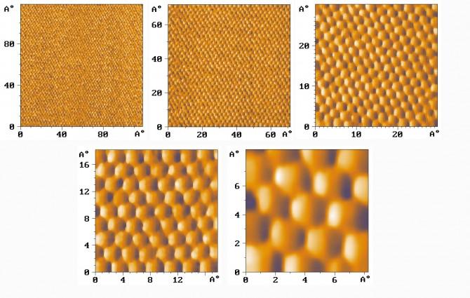
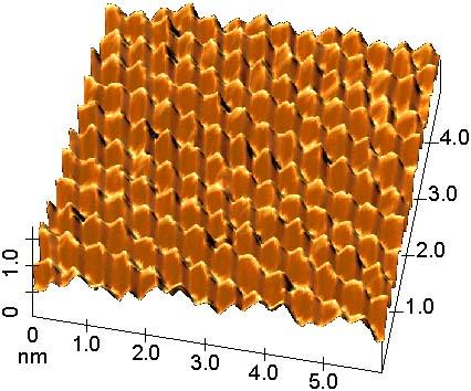
AFM
images of the highly oriented phyrolytic graphite (HOPG)
obtained in normal conditions. |
|||||||||
|
|
|||||||||
|
|||||||||
|
SFM
image of Mica atomic lattice. |
|||||||||
|
|||||||||
|
|||||||||
|
|
|||||||||
|
|||||||||
|
CaF2
epitaxial film grown on silicon(100). |
|||||||||
|
|
|||||||||
|
|||||||||
|
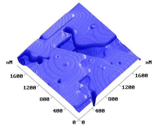
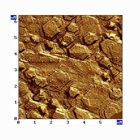
SFM
image of epitaxial MnAs ferromagnetic film grown on Si(111) with
As buffer laeyer. |
|
|
||||||||||
|
||||||||||
|
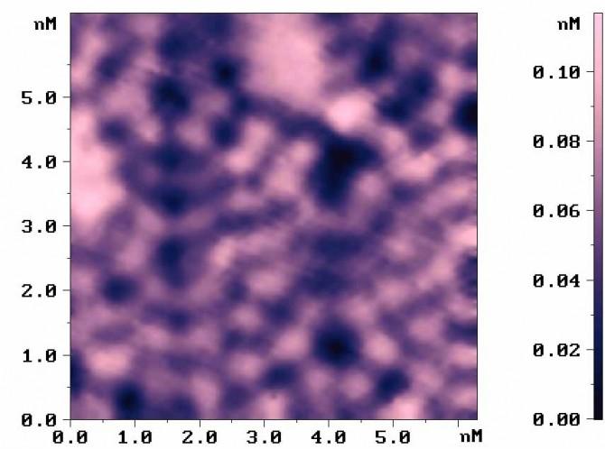
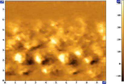
STS
image of fullerene C60 in surfactait matrix on the surface of
graphite. |
||||||||||
|
|
||||||||||
|
||||||||||
|
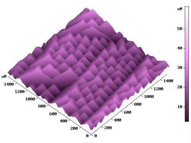
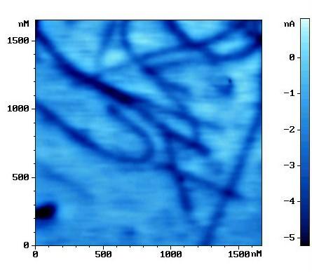
The
electrostatic force microscopy image of carbon nanotubes.
Scan size is 1,7 um. |
||||||||||
|
|
||||||||||
|
||||||||||
|
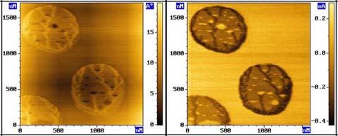
Adsorption
(most probably on the electric domain) on TGS. Left -topography;
right - phase image. |
||||||||||
|
|
||||||||||
|
||||||||||
|
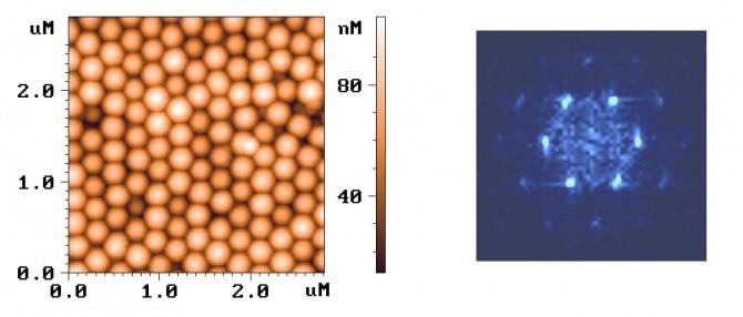
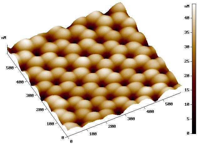
AFM image of opal matrix consisting of 200 nm SiO2 balls deposed on Si surface and central part of Fourier transform image showing good hexagonal ordering. Opal matrixes can be used as a calibration structures for SPM in submicron range covering a "dead zone" between micron range micromachined and atomic range crystal structures used for calibration now. Image courtesy of Dr. B. M. Medvedev, NT-MDT Co. |
||||||||||
|
|
||||||||||
|
||||||||||
|
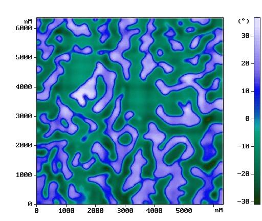
Fe-based metallic glass. The AC MFM image was obtained by two-pass technique. Image courtesy of Teodor Breczko, Physics of Metals and Nanotechnology Lab., The University of Warmia and Mazury in Olsztyn, Poland. |
||||||||||
|
|
||||||||||
|
||||||||||
|
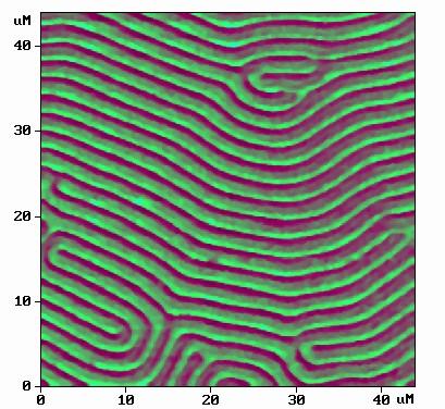
Magnetic
force derivative distribution (phase shift) of garnet film. The
period of stripe domain structure is about 2mkm. Image was
obtained by two-pass technique. |
||||||||||
|
|
||||||||||
|
||||||||||
|
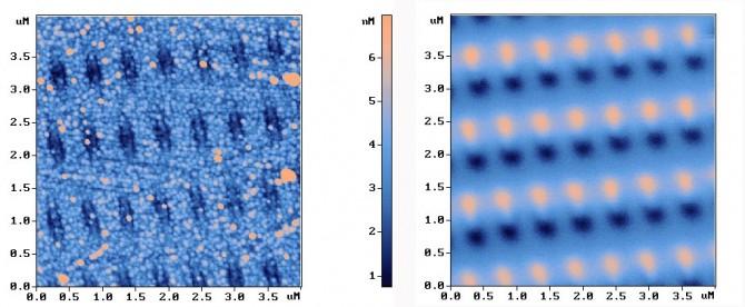
Topography
(left) and magnetic force microscopy image (right) of
ferromagnetic islands in paramagnetic film. Magnetic image looks
like a sub-micron dipole array. Dark and light areas on right
image correspond to different poles. This is perspective data
storage medium. Both images were obtained simultaneously by
two-pass method. |
||||||||||
|
|
||||||||||
|
||||||||||
|
Magnetic
bits in magneto-optical disc. Image was obtained by two-pass
technique. |
||||||||||
|
|
||||||||||
|
||||||||||
|
MFM
image of high density (40 Gb) hard disk drive. Distance between
magnetic bits is about 200 nm. MFM measurements were performed
with special magnetically soft cantilever with permalloy
coating. |
||||||||||
|
|
||||||||||
|
||||||||||
|
AFM
image of CD Al cover of replicated disk. Roughness on Top
(Sa)=3,530 nm. Roughness on Bottom (Sa)=4,055 nm. |
||||||||||
|
|
||||||||||
|
||||||||||
|
AFM
image of DVD stamper. |
||||||||||
|
|
||||||||||
|
||||||||||
|
SFM
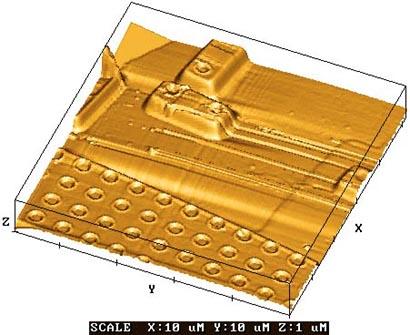
image of the surface of integrated circuit. |
||||||||||
|
|
||||||||||
|
||||||||||
|
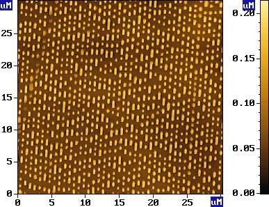
InAs quantum dots on [1,-1,0] orientation InP wafer. The sample was kindly given by Pr. Mats-Erik Pistol. Image courtesy of Sergey Lemeshko. |
||||||||||
|
|
||||||||||
|
||||||||||
|
Topography image of the self-organized matrix of anodic porous Al2O3 Images were obtained by Dr. Dmitry Kozodaev, NT-MDT. |
||||||||||
|
|
||||||||||
|
||||||||||
|
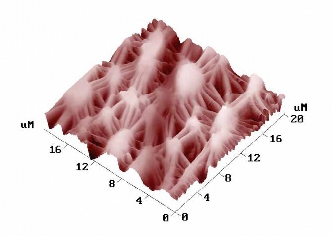
Self organized structure on the CdHgTe film surface grown by MBE Image and sample courtesy Sabinina I.V., Sheglov D.V., Sidorov Yu.G., Latyshev A.V., Institute of Semiconductor Physics SB RAS, Novosibirsk, Russia. |
||||||||||
|
|
||||||||||
|
||||||||||
|
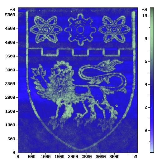
AFM nanolithography image obtained by electrical local probe oxidation technique on GaAs surface. Image courtesy of Eugeny Kuznetsov , NT-MDT, Moscow, Russia, kuznetsov@ntmdt.ru |
||||||||||
|
|
||||||||||
|
||||||||||
|
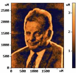
Portrait
of Zhores Alferov, russian Nobel prize winner in Physics, 2000.
AFM nanolithography image obtained by electrical local probe
|
||||||||||
|
|
||||||||||
|
||||||||||
|
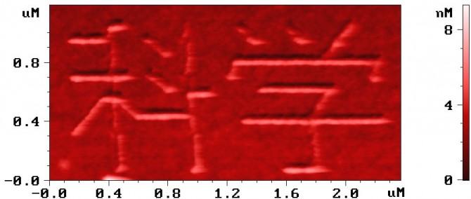
Resonance SPM modification of polycyanoacrylate film on silicon. Word "Science" in Chineese. |
||||||||||
|
|
||||||||||
|
||||||||||
|
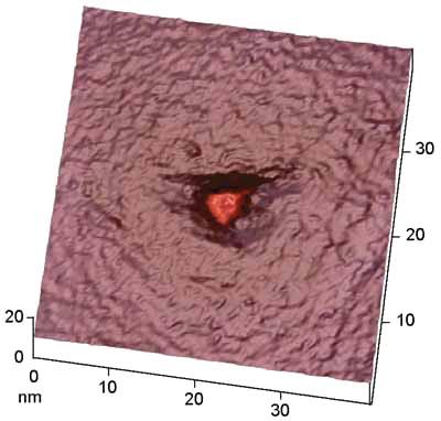
STM
image of the same LB film after local exposure to the electric
pulse with the smaller tip-surface separation (t=15mcs, a=5V,
s=0.5nm). Well-like defect appearance indicates the other
mechanism of surface modification - insulating region formation.
LB film was formed by MDT-LB5. |
||||||||||
|
|
||||||||||
|
||||||||||
|
AFM
image of the surface of fluoroplastic implantant used for
arterial |
||||||||||
|
|
||||||||||
|
||||||||||
|
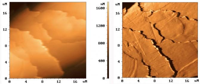
Human
hair. The figure at left is the topography, the figure at right
is the feed-back error image. |
||||||||||
|
|
||||||||||
|
||||||||||
|
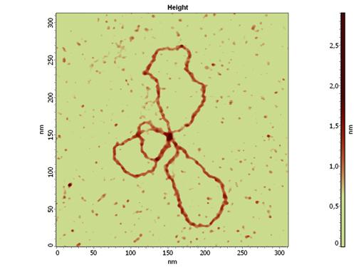
Supercoiled circular plasmide DNA (pEGFP, 3.4 kb. p.) on mica. To bind molecules to mica substrate buffer solution contains 5 mM HEPES, 10 mM MgCl2 was used. Spatial pattern of the molecule is typical for low salt buffers.The image was obtained in semicontact mode in ethanol. Triangular silicon nitride probe with spring constant 0.5 N/m was used.
Image
courtesy of M.Savvateev,
NT-MDT, Moscow, Russia, |
||||||||||
|
|
||||||||||
|
||||||||||
|
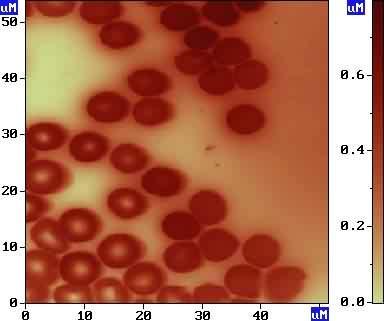
AFM
image of erythrocytes (ground-squirrel's blood). |
||||||||||
|
|
||||||||||
|
||||||||||
|
AFM
image of erythrocytes (ground-squirrel's blood). |
||||||||||
|
|
||||||||||
|
||||||||||
|
The lymphocytes of human blood fixed with glutaraldehyde on glass. Image courtesy Yu.Yu.Gushina and S.N. Pleskova, Research and Euucation Center of Solid State Nanostructures, University of Nizhny Novgorod, Russia. |
||||||||||
|
|
||||||||||
|
||||||||||
|
Mouse
fibroblast, line 3T3, fixed by formalin solution. Image obtained
by semicontact mode in liquid (phosphate buffer
solution). |
||||||||||
|
|
||||||||||
|
||||||||||
|
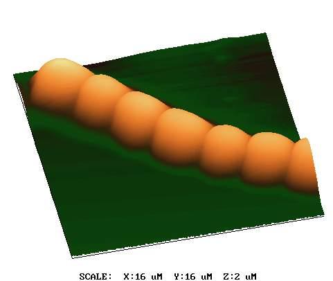
Anabaena
variabilis (ATCC 29413) cells. Cells were attached to glass
treated with L-polylysine and then fixed by 4%
paraformaldehyde. Cells were kindly given by Prof. Samuilov V.D., Moscow State University. |
||||||||||
|
|
||||||||||
|
||||||||||
|
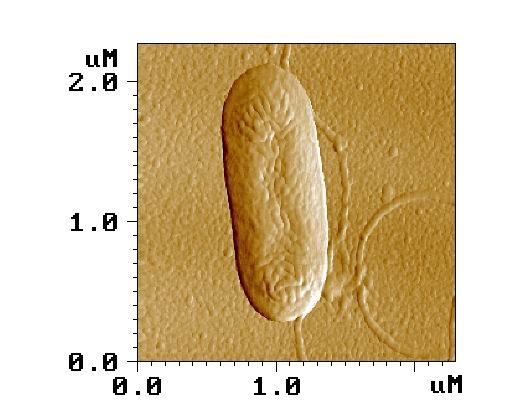
LFM
image of pseudomonas bacteria obtained in air. |
||||||||||
|
|
||||||||||
|
||||||||||
|
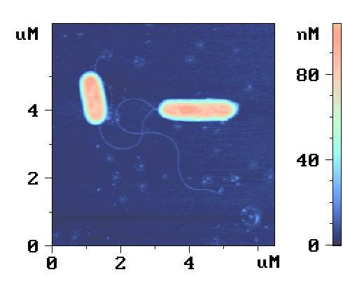
AFM
image of pseudomonas bacteria obtained in air. |
||||||||||
|
|
||||||||||
|
||||||||||
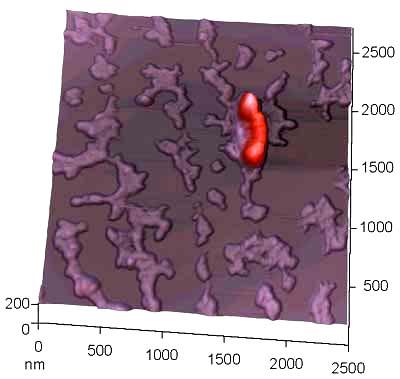
|
AFM
image of Rickettsia. |
||||||||||
|
|
||||||||||
|
||||||||||
|
Ebola
viruses inactivated in paraformaldehide solution. |
||||||||||
|
|
||||||||||
|
||||||||||
|
STM
image of Parvo Virus on HOPG. |
||||||||||
|
|
||||||||||
|
||||||||||
|
Tp
17 membrane protein net being used for syphilis
immunoassays. |
||||||||||
|
|
||||||||||
|
||||||||||
|
Monoclonal
antibodies 1RK2 to A-chain of ricin (IgG1). The characteristic
Y-shaped domain structure of the antibodies is clearly resolved.
Image obtained in semicontact mode in air. |
||||||||||
|
|
||||||||||
|
||||||||||
|
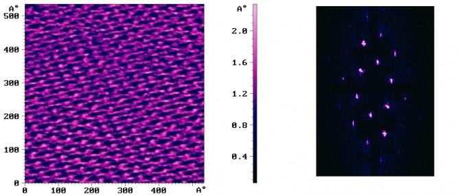
Purple
membrane imaged in liquid. Left figure is topography, right
figure is Fourier image. |
22
